The plastic component (polyethylene) in traditional hip prostheses are worn out in time and have to be replaced. With polyethylene analysis it is possible to monitor the wear in retrospective clinical studies when post-operative and end-point follow-up x-rays are available.
Typically we use AP x-rays, which make it possible to analyze linear (two-dimensionally) wear (millimeter) and a linear wear rate (millimeters per year). Prognostically, a wear rate of more than 0.2 millimeters per year shows that the polyethylene has been worn out too quickly.
By use of both AP and LA x-rays of the hip it is possible to analyse volumetric (three-dimensional) wear (mm3) and a three-dimensional wear rate (mm3 per year). In practice, LA x-rays of the hip are often poor and reduces the reproduction possiblity and the accuracy of the method.
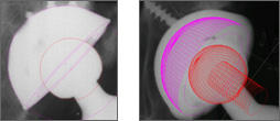
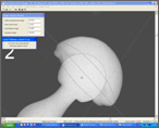
 Traditional wear analysis is done by determining two-dimensional caput femoris penetration in the polyethylen liner by use of series of digitalized x-ray images. The software PolyWare Digital Version http://www.draftware.com/pw/index.htm (Peter Devane) allows us to fit circles on the surface of caput femoris and the cup by means of a digital edge-detection algorithm. Afterwards, the software generates a three-dimensional solid model of the acetabular component and caput femoris based on the x-ray's ”back-projection” and computer-assisted design knowledge (CAD model) of the prosthesis components. Traditional wear analysis is done by determining two-dimensional caput femoris penetration in the polyethylen liner by use of series of digitalized x-ray images. The software PolyWare Digital Version http://www.draftware.com/pw/index.htm (Peter Devane) allows us to fit circles on the surface of caput femoris and the cup by means of a digital edge-detection algorithm. Afterwards, the software generates a three-dimensional solid model of the acetabular component and caput femoris based on the x-ray's ”back-projection” and computer-assisted design knowledge (CAD model) of the prosthesis components.
With Polyware Digital Version the precision of linear wear analysis is better than 0.15 mm. The quality of the x-rays has great impact on the precision of the wear analysis. If the x-rays are poor/blurred it is often necessary to set aside the digital edge-detection based circles and place them manually. This reduces the possibility of reproduction considerably.
 Wear analysis of hip prostheses can also be done by use of digitalized x-rays of the pelvis using the software Hip Analysis Suite (John Martell). The principle of the analysis is similar to the PolyWare based on edge-detection of caput femoris and the cup's surface calculating the centre. The calculation of the maximum wear is determined from the position of the centre of caput femoris and the cup on the post-operative x-ray and the last follow-up x-ray. Wear analysis of hip prostheses can also be done by use of digitalized x-rays of the pelvis using the software Hip Analysis Suite (John Martell). The principle of the analysis is similar to the PolyWare based on edge-detection of caput femoris and the cup's surface calculating the centre. The calculation of the maximum wear is determined from the position of the centre of caput femoris and the cup on the post-operative x-ray and the last follow-up x-ray.
|