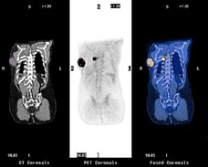
 PET scanning is based on the measurement of the decay of radioactive substances. By means of these substances it is possible to measure the flow through of blod in the bone tissue. PET scanning is based on the measurement of the decay of radioactive substances. By means of these substances it is possible to measure the flow through of blod in the bone tissue.
The technique demands execution of a conventional x-ray of the examined bone area.
Subsequently, contrast material is injected into a vein and the flow through of blod in the selected area is pictured by use of a PET scan. Then, another trace element is injected and the bone construction in the selected area is pictured with a new scan.
During the scan we take blod samples which are used in calculation algorithms. The scan takes approx. two hours.
The x-ray impact is quiet large compared to the other imaging techniques, as a scan causes 1.5 times the normal annual background radiation.
In a clinical study we have completed PET scans of the dysplastic hip joint before and after Ganz osteotomy and compared the flow through of blod in the bone. The study can contribute knowledge reducing the risk of complications and improving the results of the operations.
The PET technique could profitably be applied to several other surgical joint preservation tecniques such as hip fractures in order to reduce the risk of complications and improve the results. |